
Our goal at the Nature Center is to get people out-of-doors to see the diversity around them, to become connected to nature. Our cell phone, computers, TVs and other electronic devices have taken a hold upon us and our children to the point we remain inside and ignore the out-side world.
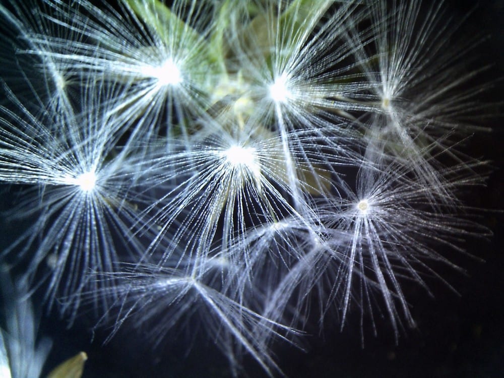
However, there is one piece of technology at the Nature Center that holds the fascination of everyone, child and adult. It is the digital microscope. Every time I walk into the Center, I see a child or adult slipping a rock, piece of wood, snake skin or other item onto the base to magnify. For me, that microscope and my digital hand lens have been the window into the microscopic world of flowers and an opening to the diversity and busyness of that world not visible to the human eye.
Craig Martin and I just finished teaching a class entitled “How to Identify Wildflowers.” We are revising the book Dorothy Hoard and I wrote some years ago. Dorothy and I were just beginning the revision when she died. Dorothy originally had done over 400 drawings, an amazing monumental task. My task, for our new project, is detailed drawings of flower parts, leaf, stem, and textures. With the hand lens and the digital microscope I can see the detail I want to put into the drawing.
In botany, we have long used microscopes and hand lenses (magnifying glasses) to see detail. We have even been able to take pictures. However, in recent years digital hand lenses have been on the market. They not only magnify but allow easy picture-taking of the magnification. As part of my arsenal for flower identification, I purchased such a hand lens.
When I first put a flower under the hand lens, I saw a small aphid peeking over the base of the flower, others scampering about busily, perhaps unaware their world had been disrupted and discovered. With each new flower, leaf, or stem I saw the microscopic world magnified: glandular hairs, star-like hairs, unusual shapes, tiny insects. It was like walking into one of our canyons, along a trail and observing the diverse world of birds, flowers, rocks, and textures. It is a fascinating and amazing microscopic world.
A regular dissecting scope is usually a one-person experience. You can look in the scope, find and item and have one person look at it. This year, I used the digital microscope in the class. The advantage was that the students could gather around the screen. We could simultaneously view an item, discuss it, and, Ohoo and awe at the unique structures of each individual plant. We not only were able to see the amazing diverse world we live in by our field trips to various plant communities, but with this tool, we were also able to look into the amazing microscopic world of flowers.


